Avian Skeletal System
 Prepared for the 2012 MacFarlane Pheasant Symposium
By: Rob Porter, DVM, PhD
Minnesota Veterinary Diagnostic Laboratory
How does avian skeleton differ from mammalian skeleton?
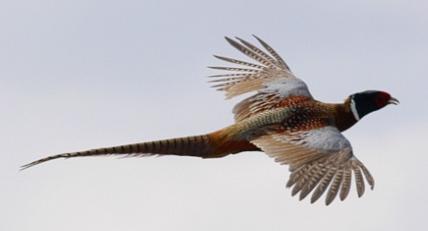
When animals adapt to particular environments their physical structures will change (natural selection) to take advantage of that environment (eating and reproduction). For example, humans are adapted to walking and running with an upright skeletal structure. Birds are adapted to flight and that is reflected in the structure of the skeleton and individual bones.
Ossification
Bird bones are relatively thin, but are stiff and dense compared to mammal bone. One adaptation is fusion of vertebrae to form a rigid spinal column to support flight.
Bone mass
Birds have reduced absolute bone mass compared to mammals, but the avian bone is generally more dense, indicating a higher strength-to-weight ratio in birds.
Pneumatic bone
Avian bones are often pneumatic (hollow; infiltrated by air sacs). Pneumatic bone is much more extensive in birds that fly for long distances, e.g. songbirds. Penguins and ostriches are ground dwelling birds that have very little pneumatic bone. Gallinaceous birds, such as chickens, turkeys, pheasants, and partridges, have a moderate amount of pneumatic bone. Pneumatic bone has a honeycomb structure on cross-section. The medullary cavity of pneumatic bone in the avian skeleton contains diverticula of internal air sacs. In the chicken, and likely other gallinaceous birds, the sternum, humerus, pelvis, cervical and thoracic vertebrae are pneumatic.

Cross-section of pneumatic vertebral bone reveals lacey, interconnected air channels and cross-branching trabeculae for bone support.
Bone Function
The skeletal system consists of bone and has the following functions:
- Protection and support of internal organs, e.g. heart and lungs
- Source of calcium for egg production
- Articulated, jointed system for movement- walking and flight
- Supportive matrix for stem cells that form cells of the peripheral blood- granulopoiesis(leukocytes) and hematopoiesis (red blood cells)
Bone Composition
Inorganic matrix: Bone mineral content = bone ash is approximately 55-65% of bone by dry weight. Consists of hydroxyapatite, which has the chemical formula Ca5(PO4)3(OH) and is a crystal produced by calcium and phosphorus. It is the major component of bones and teeth and provides strength and hardness. Organic matrix (35%) = protein which is mostly type 1 collagen, which gives bone flexibility.

Skeletal Anatomy
The skeleton can be divided into axial and appendicular segments:
- Axial skeleton: central axis of body = skull, vertebral column, ribs and sternum
- Appendicular skeleton: Pectoral and pelvic girdles, wings and legs
Skull
Only few bones in the skull are movable. These are the lower jaw bones, roof of the mouth and bones of the tongue. Orbits encase the eyes and are large. Ridges or hard edges of the beak substitute for teeth.

Skull (chicken): Note the scleral ossicle (arrow), which is a ring of bone that surrounds and supports the eyeball within the orbit.
Vertebral (spinal) column
Provides base for the bones of the trunk and limbs and is main support for skull. Spinal column is composed of chain of bones referred to as vertebrae. These vertebrae provide passage and protection for the spinal cord and nerve roots.
- Cervical vertebrae (C) – extend from the skull to the first thoracic vertebral body
- Thoracic vertebrae (T) – each is connected to two ribs, one on each side. The first three thoracic vertebrae are fused in chickens.
- Lumbar and sacral vertebrae (L and S) – these are fused together into one long synsacrum that also includes several fused caudal vertebrae.
- Caudal vertebrae: The last 3-4 caudal vertebrae are fused into single, flattened bone called the pygostyle, which provides attachment for several tail feathers (rectrices).


Oval outlines the fused thoracic vertebrae that give the spine rigidity for flight
Ribs
A flattened arch of bone that is attached to the last cervical or thoracic vertebrae and the sternum. Uncinate processes project from one rib to an adjacent rib to form a firm ribcage.
Thoracic ribs (arrows) have uncinate processes (stars), which are projections overlapping and fused to adjacent ribs to create a more rigid thoracic cage.


Sternum
Breastbone is flattened in birds that do not fly, e.g. ostrich. Most birds, including gallinaceous birds, have a carinate or keeled sternum that resembles the keel of a boat. This provides greater surface area for the flight muscles to attach.
Sternum of gallinaceous birds has a broad, flat, but narrow surface for attachment of flight muscles on either side.
Pectoral Girdle
The pectoral or shoulder girdle consists of three bones (“tripod”) to support the wing bones. The girdle consists of 1) coracoid bone, 2) scapula and 3) furcula = clavicle. The coracoid and scapula form a pocket (glenoid cavity) for the head of the humerus to attach. The coracoids, scapula and calvicle also form the foramen triosseum, which is a channel for the tendon of an important flight muscle, the supracoracoideus muscle.
Sternum of gallinaceous birds has a broad, flat, but narrow surface for attachment of flight muscles on either side.

Skeleton
The bones of the pelvic girdle are the ilium, ischium and pubis. These provide a socket (acetabulum) for the head of the femur.

Skeleton
The space between the ends of the pubic bones are greatest in birds that are currently in egg production.
Wing
The skeleton of the wing consists of the humerus, radius and ulna, carpal bones, a carpometacarpus and digits. The humerus lies close to the thoracic cavity when the bird is at rest and connects with the glenoid cavity of the pectoral girdle. Tendons of two large breast muscles, the supracoracoideus and superficial pectoral muscle, attach to opposite sides of the humerus to raise and lower the wing during flight.

Leg
The pelvis limb is composed of femur, patella, tibiotarsus, fibula, tarsometatarsus, metatarsal bone and four digits. The tarsometatarsus represents fusion of a distal row of tarsal bones with the three metatarsal bones of digits II, III and IV.

Gallinaceous birds such as pheasants and partridges have feet with three digits pointing forward (craniad) and one digit pointing backward (caudad).

Major bones of the galliform leg (turkey).

Anatomy of tibiotarsus = drumstick. Bone formation largely at the metaphysis (growth plate) at each end of long bone causes marked elongation (dotted lines) of the bone during the bird's growth phase.

Cross-section of diaphysis (shaft) of tibiotarsus (drumstick).

Bone ash represents portion of dry bone that is composed of mineral.
Tibial Rotation and Valgus Deformation (Splay leg, slipped tendon)
Definition
A medial rotation and angulation, usually of the of the tibiotarsal bone in one of both legs, resulting in recumbency, lameness, and a splay-leg appearance. This occurs in young birds and results in permanent lameness.
Cause
Usually unknown cause. This occurs in one or both of the legs mostly in young birds with rapidly growing bones, particularly the tibiotarsus. I suspect that the cause of the abnormal growth can be multifactorial and includes a 1) congenital predisposition based on inherited traits, 2) early metabolic effects on bone growth caused by decreased vitamin D3 or abnormal ratio of calcium and phosphorus in the ration, and 3) poor traction or footing in the pen (smooth surface or wet, slick litter on floor). In addition, any leg injury that causes the bird to favor or alter the stresses placed on one leg can result in abnormal growth, especially during the stages of rapid bone formation and elongation.


Four-week-old white pheasants: Abnormal posture and recumbent birds caused by deformation of one leg.

Left hock (intertarsal) joints of abnormal and normal 4-week-old white pheasants. In the abnormal hock joint the gastrocnemius tendon (solid line) has slipped laterally and outside of the groove (marked by dotted lines) within the distal tibiotarsus.

Four-week-old pheasants: Normal and abnormal left tibiotarsal bones (anterior aspect); In the abnormal bone note the bowing of the proximal shaft (diaphysis) and inward (medial) angulation of the proximal metaphysis (Photo A) and a slight rotation of the proximal epiphysis (articular surface) in the abnormal bone (Photo B).

Clinical Signs
Because of the rapid growth rate of a young bird the signs of lameness can appear quickly, usually only affecting a small percentage of the flock. The skin of the hock joint can be dark red or scabbed as a result of the bird using this surface rather than the foot to support the body. Birds that are severely affected will be recumbent with poor uniformity because of an inability to compete. The tarsometatarsus often projects laterally (outward) in a valgus (“knock-kneed”)deformation because of the angle formed at the proximal tibiotarsal bone. In my experience the femur and tarsometatarsus usually appear normal.
Gross Lesions
In pheasants the affected leg often projects laterally (outward) because of the angle formed in the proximal tibiotarsal bone at the junction of the diaphysis and metaphysis. This bone can be bowed at the proximal aspect. The femur and the tarsometatarsus can be normal. The angulation of the proximal tibiotarsus increases the tension on the gastrocnemius tendon, which can slip out of the articular groove in the distal portion of the bone (see photograph). The displaced tendon is secondary to the abnormal bone growth, and the extended leg often cannot be easily flexed.
Treatment/Prevention
There is no immediate treatment for the affected limb and the emphasis should be prevention. Maintain dry flooring with good traction in the pen and closely monitor the nutrients that will affect bone growth, particularly vitamin D3, Ca, P and trace minerals. Breeder flocks that produce a greater percentage of affected chicks should be avoided if possible.
Also referred to as rickets in young birds, osteomalacia is decreased mineral in bone resulting in soft, pliable bones including the beak, curved keel bone and beading of the ribs at the costochondral junction. Osteomalacia is diagnosed most often in young, growing birds and hens in active egg production. Affected birds can be found on their sides in the back of the cage. At the time of initial paralysis, birds can appear healthy and will have a shelled egg in the oviduct. Death occurs from starvation or dehydration, a failure of the birds to reach the feet or water. A high incidence of osteomalacia can be prevented if proper ratios of calcium and phosphorus in the diet are maintained. Soft bones occurring in young birds is referred to as rickets. This disorder is usually caused by abnormal Ca/P ratios in the ration, low vitamin D3 levels in the ration, or intestinal malabsorption associated with enteric diseases. The birds are often recumbent, but bright and alert with extremely pliable beaks and long bones.

Laying Hen With Deformed Keelbone
Osteomalacia: Laying hen with deformed (Sshaped) keelbone

Turkey with Rickets
Rickets: Nonambulatory turkey poult
Further Reading
Aves Introduction, by AS King, In Sisson and Grossman’s The anatomy of Domestic Animals, R Getty (ed.), Fifth Ed., W.B. Saunders Publishing, Philadelphia, PA, 1975.
Birds Their Structure and Function. AS King and J McClelland (eds.), Bailliere Tindall, London, 1977.
E R Dumont. Bone density and the lightweight skeletons of birds. Proc. Royal Soc. B , 277:2193-2198, 2010.
Manual of Ornithology, Avian Structure and Function. NS Procor and PJ Lynch (eds.) Edwards Brothers, Inc. Ann Arbor, MI, 1993.
Ornithology in Laboratory and Field, Fourth Edition, O S Pettingill, Jr. (ed.), Burgess Publishing Co., Minneapolis, MN, 1975.
Sturkie’s Avian Physiology, Fifth Edition. GC Causey (ed.). Academic Press, San Diego, CA. 2000.